What is it about?
Intraoperative Doppler ultrasonography is a noninva- sive imaging modality that provides an unrestricted real- time method for identifying the vertebral artery (VA) and quantifying its blood flow during C1–C2 posterior arthrodesis. It may represent a useful additional tool supporting fluoroscopy-assisted procedures, but it may also play a significant role even during surgeries in which neuronavigation is used, reducing the chance of a mismatch between the view on the neuronavigation screen and the actual course of the VA in the operative field and providing the adjunctive data of the VA’s blood flow. Doppler ultrasonography is an easy and economical modality that may help prevent vertebral artery injury (VAI). Since this surgical tool is already used in neurovascular procedures, there is no learning curve. Moreover, the possibility of estimating the blood flow velocity before and after arthrodesis, intraoperatively verifying the impact of surgery through any eventual impairment of blood flow values, may help in the early detection of subtle complications, such as VA pseudoaneurysms and stenosis, thus providing an advantage in terms of patient outcome.
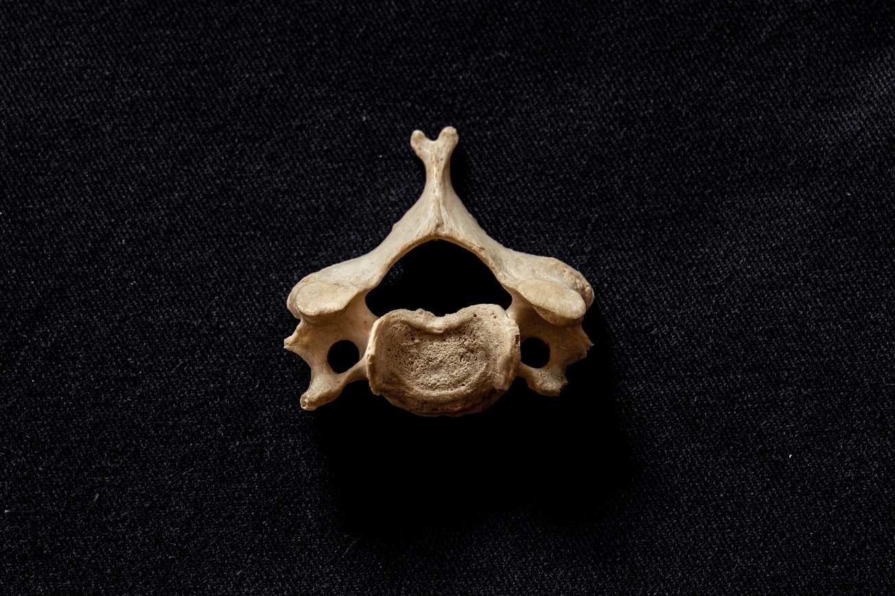
Featured Image

Photo by Meta Zahren on Unsplash
Why is it important?
Intraoperative doppler ultrasound is an incredible tool to increase safety of C1-C2 fixations
Read the Original
This page is a summary of: Intraoperative Doppler ultrasound as a means of preventing vertebral artery injury during Goel and Harms C1–C2 posterior arthrodesis: technical note, Journal of Neurosurgery Spine, December 2019, Journal of Neurosurgery Publishing Group (JNSPG),
DOI: 10.3171/2019.5.spine1959.
You can read the full text:
Resources
Contributors
The following have contributed to this page


