What is it about?
We measured anomalous diffusion in human prostate cancer cells which were transfected with the Alexa633 fluorescent RNA probe and co-transfected with enhanced green fluorescent protein-labeled argonaute2 protein by laser scanning microscopy. The image analysis arose from diffusion based on a “two-level system”. A trap was an interaction site where the diffusive motion was slowed down. Anomalous subdiffusive spreading occurred at cellular traps. The cellular traps were not immobile. We showed how the novel analysis method of imaging data resulted in new information about the number of traps in the crowded and heterogeneous environment of a single human prostate cancer cell. The imaging data were consistent with and explained by our modern ideas of anomalous diffusion of mixed origins in live cells. Our original research presented in this study is significant as we obtained a complex diffusion mechanism in live single cells.
Featured Image

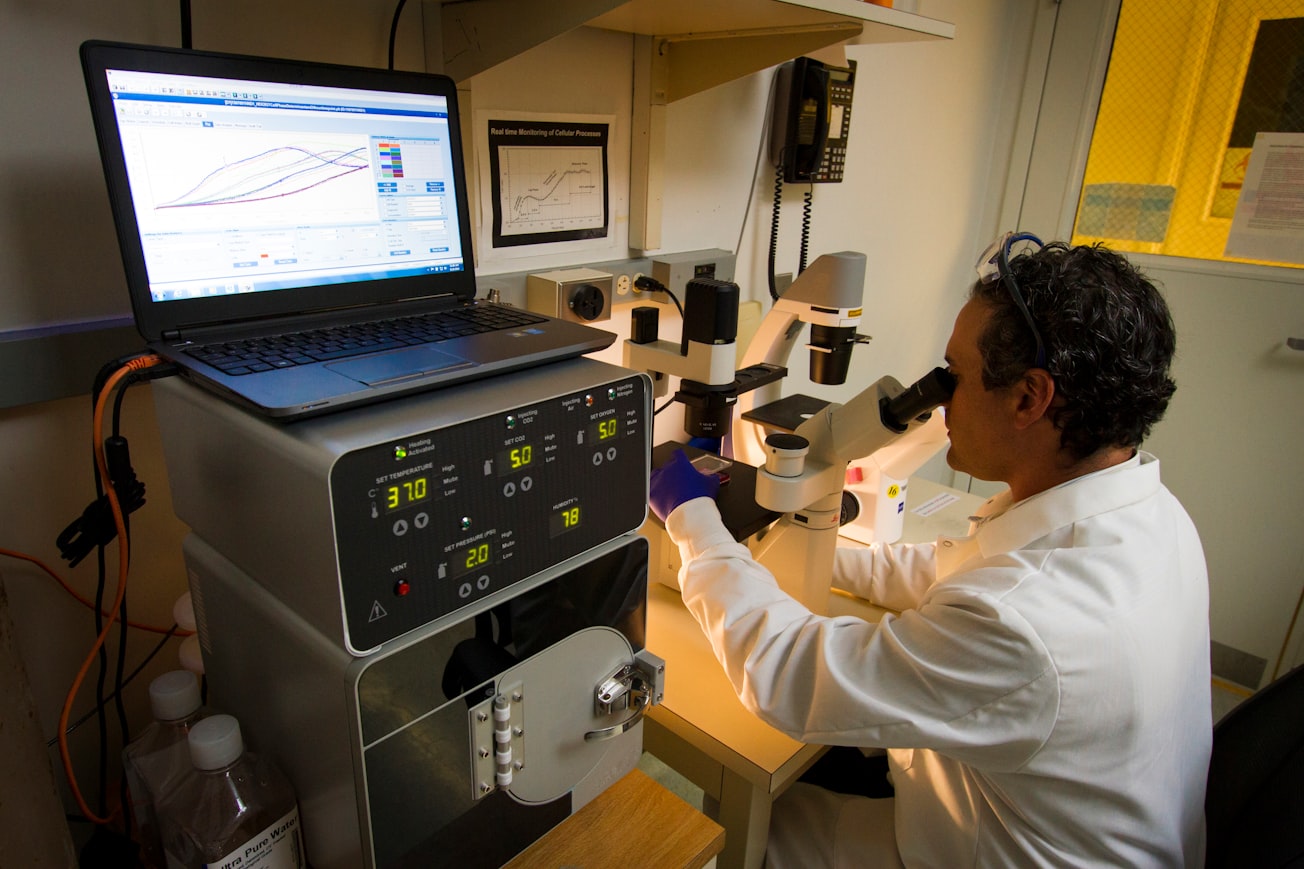
Photo by National Cancer Institute on Unsplash
Why is it important?
The original paper described FCS studies on an Alexa633 labeled RNA probe and green fluorescent protein-labeled argonaute2 protein in human prostate cancer cells. An interesting and originally “twist” to the experiments was the use of a so-called superquencher, BHQ3, which is not fluorescent, but which quenches the Alexa633 labeled RNA when it is in the duplex form. When the RNA strands dissociate upon binding to the EGFP-argonaute2 protein the Alexa633 then fluoresces brightly. We investigated human PC-3 cells expressing EGFP-Ago2 and transfected with the Alexa633-tagged dsRNA probe. Our mathematical approach, fully described in the text, was a convolution of exponential functions in form of the gamma distribution density. The method assumed that traps were not immobile and was able to extract their numbers in crowded and heterogeneous environment of living cells. Using our novel analysis method of imaging data allowed us to obtain new information about the number of traps in a crowded and heterogeneous cellular environment. The theory and experimental approach, using two different laser wavelengths, were described in sufficient detail to allow others to replicate their findings and apply to approach to other biological systems. The work appears to be a highly original and significant addition to the growing armamentarium of FCS and LSM techniques. The research article is an interesting paper about a complex diffusion occurring in live cells. In a crowded cell environment one can expect such complexity.
Perspectives
It is about the single-molecule level versus the many-molecule level in a single live cell. The advantages of the physical 'Theory of Single-Molecule Biophysics & Biochemistry Based On Individually Diffusing Molecules in Dilute Liquids and Live Cells' are evident: The 'SINGLE-MOLECULE DEMON' (single-molecule ratchet) in IMAGING/MICROSCOPY/SUPER-RESOLUTION MICROSCOPY (NANOSCOPY) and SPECTROSCOPY: Dilute Liquids and Live Cells •https://www.growkudos.com/publications/10.2174%252F138920111795470949/reader •https://ajtm.journals.publicknowledgeproject.org/index.php/ajtm/article/view/1408/2082 The thermodynamic Single-Molecule DEMON: How to avoid him in the measurements of dilute liquids and live cells without immobilization or flow: https://pubmed.ncbi.nlm.nih.gov/35713144/
PRESERVE FROM BEING FORGOTTEN: Professor Zeno Földes-Papp [Biochemist, Gerontologist (Biochemiker, Geriater)]: Laying the Foundation of Single-Molecule Biophysics & Biochemistry Based On the Stochastic Nature of Diffusion: The Individual Molecule, from the Mathematical Core to the Physical Theory. -- I hope that my humble scientific work will be well received by the communities of single-molecule imaging and spectroscopy and by all users of these technologies as well as biotechnologies in the various and different disciplines:
Head of Geriatric Medicine (Medical Director of the Geriatric Service: Sektionsleitung Geriatrie) at Asklepios Klinikum Lindau (Bodensee), Bavaria, Germany
Read the Original
This page is a summary of: Visualization of subdiffusive sites in a live single cell, Journal of Biological Methods, January 2021, Journal of Biological Methods,
DOI: 10.14440/jbm.2021.348.
You can read the full text:
Resources
PubMed
Research Article
Single-phase single-molecule fluorescence correlation spectroscopy (SPSM-FCS)
This information has been edited and peer-reviewed by contributors to the Natural Standard Research Collaboration (www.naturalstandard.com).
What do Single-Molecule Localization or Single-Molecule Analysis in liquids or live cells without immobilization or hydrodynamic flow tell us about a single molecule?
Have a deeper look at the Theory of Single-Molecule Detection of one and the same molecule (the individual molecule) in dilute liquids and single live cells without significant hydrodynamic flow or immobilization on artificial or biological surfaces/membranes: The Single-Molecule Time Resolution in microsopy/nanoscopy (super-resolution microscopy)/ spectroscopy: https://lnkd.in/dXwqMrV https://lnkd.in/dbZKRyg https://lnkd.in/dbE4z23 https://lnkd.in/dgegX6Y Sorry, the biggest breakthrough in microscopy/nanoscopy and spectroscopy would be a breakthrough in sensitivity for measuring an individual single molecule (one and the same molecule) over several minutes without immobilization or significant hydrodynamic flow. Interested in joining this group? Single Molecule Biophysics & Biochemistry,see https://www.researchgate.net/profile/Zeno_Foeldes-Papp
Contributors
The following have contributed to this page


