What is it about?
Nonmetallic inclusion (NMI) populations in superelastic (SE) Nitinol fine wires are critical to the understanding of the wire's fatigue behavior. Characterization of NMIs, particularly in very fine wires (less than 150 microns in diameter) is challenging. This investigation combined plasma focused ion beam (PFIB) serial sectioning with scanning electron microscopy (SEM) to provide qualitative and quantitative information about the NMI populations. This technique resulted in three-dimensional (3D) reconstructions providing a more complete connectivity of NMIs and pores as well as information about the distribution of the features within the wire volume that is not possible with traditional two-dimensional (2D) imaging techniques.
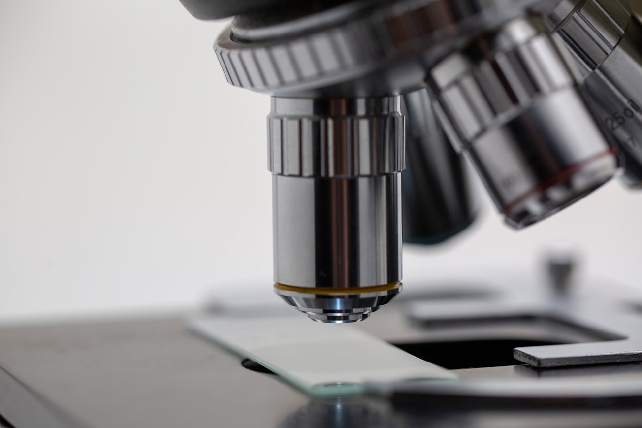
Featured Image

Photo by Michael Longmire on Unsplash
Why is it important?
Many biomedical devices and medical tools are constructed from Nitinol wire or thin-walled tubing that demand long-term reliability. Studies have shown that fatigue failure is correlated with nonmetallic inclusion populations. The ability to more accurately characterize these features will allow for the development of more robust fatigue modules and provide valuable input for Nitinol melt process optimization.
Perspectives
While traditional metallographic approaches are the cornerstone for characterizing nonmetallic inclusions, the 3D connectivity of the features is lost. PFIB-SEM serial sectioning is a valuable tool to provide an enhanced understanding of the NMI morphology, connectivity, and allows for a more accurate quantification of the population.
Janet L. Gbur
Case Western Reserve University
Read the Original
This page is a summary of: Plasma Focused Ion Beam Serial Sectioning as a Technique to Characterize Nonmetallic Inclusions in Superelastic Nitinol Fine Wires, Microscopy and Microanalysis, December 2020, Cambridge University Press,
DOI: 10.1017/s1431927620024617.
You can read the full text:
Contributors
The following have contributed to this page


